Internal geometry and density contrast
industrial CT
Material Analysis · Microscopy & Imaging
Characterise internal geometry and density contrast, pores, cracks, inclusions, interfaces, and connectivity, and three-dimensional dimensions and volume fractions using a scope agreed around your sample and decision.
Why this test
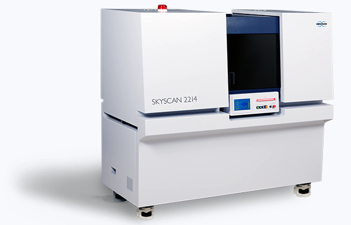
Computed Tomography (Industrial / Micro-Nano / In-situ / Synchrotron CT) is selected when the project needs defensible information on internal geometry and density contrast, pores, cracks, inclusions, interfaces, and…
industrial CT
micro- or nano-CT
region-of-interest or in-situ CT
Choose the scope
The measurement mode, preparation route and reporting depth depend on what you need to learn from the sample.
internal geometry and density contrast
pores, cracks, inclusions, interfaces, and connectivity
three-dimensional dimensions and volume fractions
Common outputs
Fields, measurement conditions, processing and file formats are confirmed before work begins.
Reconstructed slice stack reported with the agreed units, sample reference and measurement conditions.
3D volume and rendered views supplied in a labelled format suitable for direct sample or condition comparison.
Segmentation, porosity, defect, or dimensional metrics when scoped provided with the processing basis and quality checks agreed during technical review.
Method-specific images included when selected in the confirmed reporting scope.
Sample requirements
Provide representative, clearly labelled samples and identify the region, feature or comparison that matters.
| Suitable sample or input | Submission requirement | Planning note |
|---|---|---|
| Powder / particles | Provide a dry, homogeneous and clearly labelled sample in a sealed container. | Exact mass and any drying or dispersion step are confirmed after technical review. |
| Liquid / solution | Provide a sealed, compatible container labelled with composition, concentration and solvent. | Declare volatility, corrosivity, suspended solids and any storage or temperature requirement. |
| Synthetic soft material | Provide fixed, preserved or otherwise stabilised material in the agreed container and medium. | Describe the material composition and confirm preparation, temperature and disposal requirements. |
| Test coupon / specimen | Provide labelled specimens prepared to the agreed geometry, orientation and surface condition. | Confirm the applicable standard, dimensions, loading direction and number of replicates. |
Region of interest: include a marked image, sketch or reference when the target feature is localised.
Handling: declare hazardous, magnetic, volatile, moisture-sensitive, beam-sensitive or temperature-controlled material before dispatch.
Questions and answers
Short answers to issues that can change preparation, scope or interpretation.
Provide powder / particles, liquid / solution, synthetic soft material, test coupon / specimen as applicable. Exact quantity, dimensions and preparation are confirmed during technical review.
Choose the mode around the decision you need to make. For internal geometry and density contrast, start with industrial CT; add micro- or nano-CT when comparison or quantification is required.
Typical turnaround is 8–12 working days after sample acceptance and method confirmation.
Resolution trades off against sample size and attenuation. Materials with similar X-ray attenuation may be difficult to segment without contrast enhancement.
Configure the service
A £45 service fee applies to orders under £300, excluding VAT.
Typical turnaround: 8–12 working days
Configure & Book