Particles and powders
Compare particle shape, surface texture, broken particles and agglomeration.
Material Analysis · Microscopy & Imaging
High-resolution surface imaging for particles, coatings, fracture surfaces, pores, fibres and microstructural defects, with optional EDS elemental analysis.
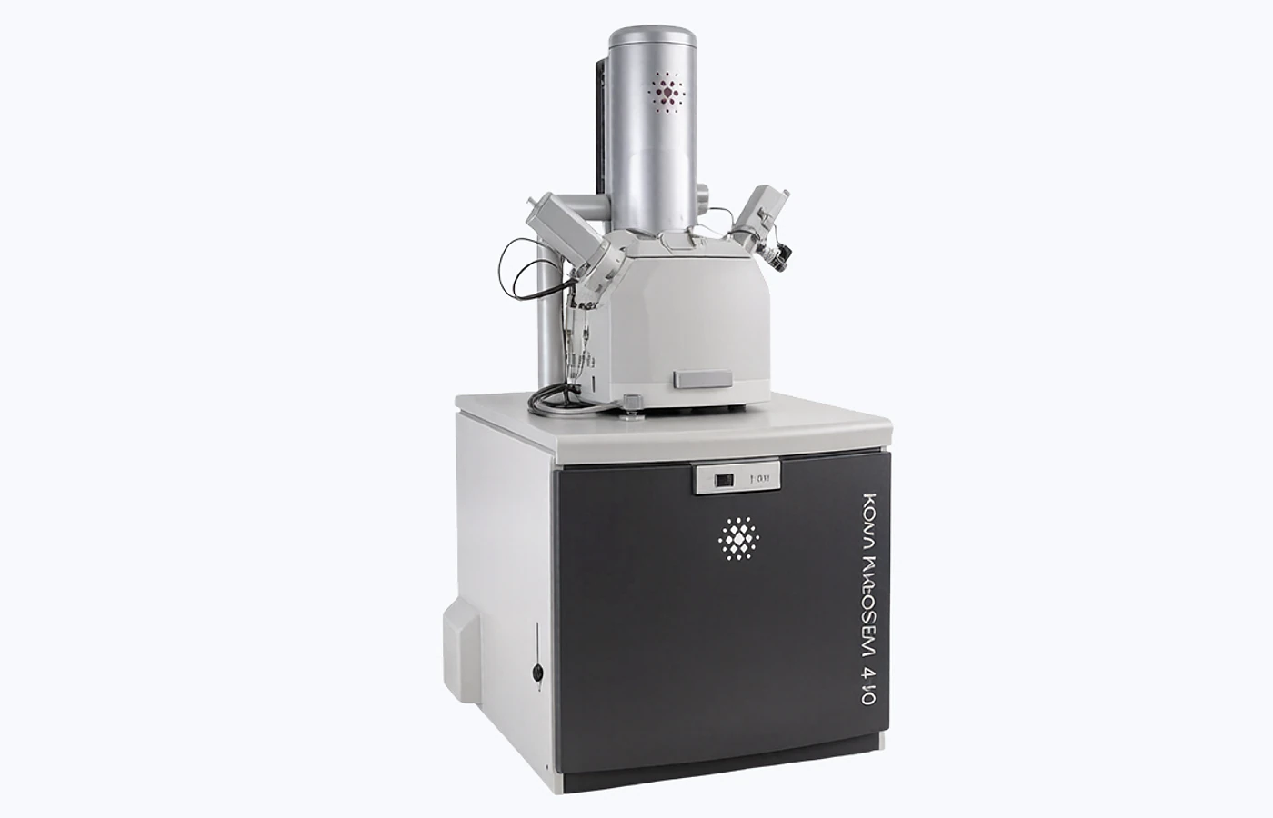
Why this test
SEM reveals surface and near-surface features from the micrometre to nanometre scale. It is most useful when morphology, local defects or composition must be tied to a specific feature.
Compare particle shape, surface texture, broken particles and agglomeration.
Inspect fracture texture, crack origins, inclusions and processing defects.
Check coverage, pinholes, cracks, delamination and interface quality.
Locate deposits, pitting, debris and surface damage linked to service conditions.
Add EDS to identify elements at selected points, paths or mapped areas.
Choose the scope
The selected instrument, detector and preparation route depend on what you need to learn from the sample.
Best for particle shape, surface texture, pores, fibres, coating coverage and fine defects.
Best for inclusions, residues, contrasting phases, coating distribution and contaminant particles.
Best for fracture origins, interfaces, buried defects, diffusion zones and layered structures.
Common outputs
Image count, magnifications, fields of view and file formats are confirmed before testing.
Calibrated SEM image set supplied with the agreed units, labels, sample reference and measurement conditions.
SE morphology images supplied with the agreed units, labels, sample reference and measurement conditions.
BSE contrast images supplied with the agreed units, labels, sample reference and measurement conditions.
EDS point spectra supplied with the agreed units, labels, sample reference and measurement conditions.
EDS line profiles supplied with the agreed units, labels, sample reference and measurement conditions.
Elemental maps supplied with the agreed units, labels, sample reference and measurement conditions.
Data files: image and EDS files, acquisition metadata and raw files are supplied where available and agreed in the quotation.
Example results

Mixed particle sizes, shapes and agglomerates collected on a PTFE filter.
Akmaral Agibayeva · source · CC BY
Microvoid coalescence on a 6061-T6 aluminium tensile fracture surface.
Bob Clemintime · source · CC BY-SA
Pearlite in eutectoid steel after annealing and Nital etching.
Edward Pleshakov · source · Public domain
High-magnification view of internal microvoids within a synthetic fibre.
Innegratech · source · CC BY-SASample requirements
Dry, clean and clearly marked samples reduce avoidable preparation time and make the returned images more useful.
| Sample type | Preferred amount or size | Preparation and information required |
|---|---|---|
| Powders | 5–10 mg | Dry and sealed. State whether particles should be imaged as received or dispersed. |
| Films and bulk solids | Up to 10 × 10 × 5 mm preferred | Mark the surface, cross-section or region of interest. Larger samples require review. |
| Liquids and pastes | Method dependent | Submit only when fully dried or when drying has been agreed before testing. |
| Cross-sections | Fit to the agreed holder | Provide a clean prepared face or request sectioning, polishing, ion milling or FIB. |
| Non-conductive samples | As above | Au/Pt is commonly used for morphology; carbon may be preferable for EDS-sensitive work. |
| Special-risk samples | Advance review required | Declare magnetic, moisture-sensitive, volatile, beam-sensitive, toxic or radioactive materials. |
Vacuum compatibility: standard high-vacuum SEM is not suitable for wet or volatile samples.
Region of interest: include a marked photograph or sketch when the target feature is localised.
Questions and answers
Short answers to the issues that most often change preparation, scope or interpretation.
No. Conductive and well-grounded samples may be imaged uncoated. Non-conductive samples often need a thin conductive coating to reduce charging.
Au/Pt usually gives strong morphology imaging. Carbon is often chosen when metal coating peaks could interfere with EDS.
SE imaging emphasises surface topography. BSE imaging is more sensitive to average atomic number and can reveal compositional or phase contrast.
Point analysis measures one location; a line profile follows change along a path; mapping shows selected element distributions across an area.
EDS is normally semi-quantitative. Results depend on preparation, coating, geometry, matrix and concentration. Hydrogen and helium are not measured and light-element sensitivity is limited.
They require advance review. Standard high-vacuum SEM is not suitable for wet or volatile samples, and magnetic samples may be restricted by the selected instrument.
Request a quote
A £45 service fee applies to orders under £300, excluding VAT.
Typical turnaround: 8–12 working days
Configure & Book